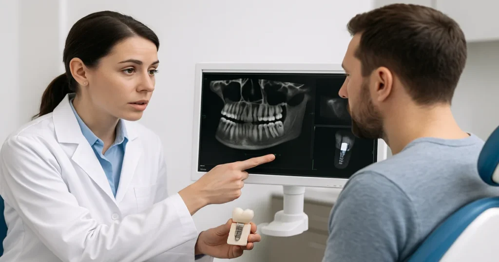
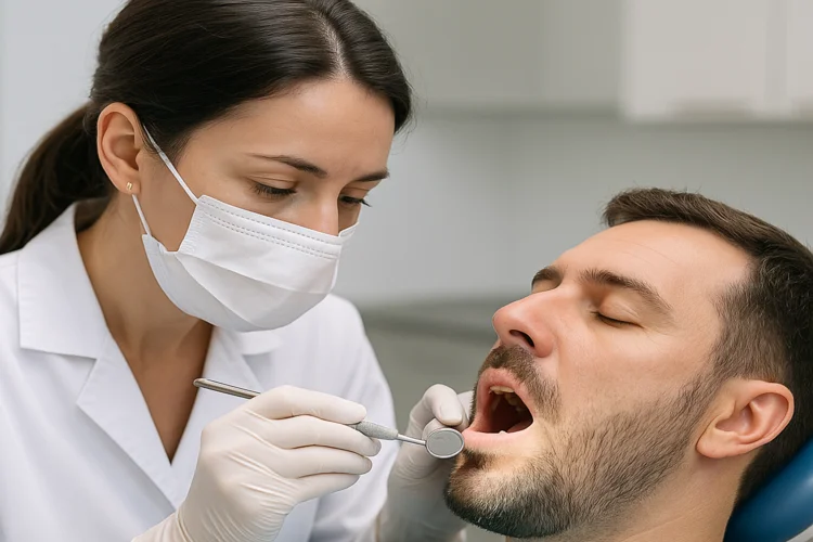
Every successful dental implant begins with accurate planning and execution. Even a minor miscalculation in bone depth or angulation can compromise stability, esthetics, or nerve integrity. That’s why modern implant dentistry relies on Cone Beam Computed Tomography (CBCT) to visualize the jaw in three dimensions.
Traditional X-rays provide limited, two-dimensional data. In contrast, a CBCT scan captures detailed information about bone structure, sinus positioning, and nerve pathways, factors essential to treatment success.
In this article, we’ll explore when to take a CBCT scan for implant placement, how timing and clinical indications affect outcomes, and the evidence-based guidelines that help dentists use this technology effectively and responsibly.
What Is CBCT and Why Does It Matter in Implant Dentistry
CBCT is a 3D imaging technology that produces highly accurate visualizations of teeth, bone, and surrounding anatomy. The scan uses a cone-shaped X-ray beam that rotates around the head, generating detailed images for diagnosis and surgical planning. Compared to medical CT scans, CBCT offers greater spatial resolution at a significantly lower radiation dose.
In implant dentistry, this technology is invaluable. It enables clinicians to assess bone density and volume, identify the course of the inferior alveolar nerve, and visualize the maxillary sinus prior to implant surgery. CBCT also supports advanced procedures, such as bone grafting, sinus lift surgery, and periodontal regeneration, where anatomical precision is crucial for clinical success.
How CBCT Improves Treatment Planning
CBCT eliminates guesswork by allowing dentists to plan implant placement digitally. Virtual 3D models enable exact measurements of implant length, diameter, and trajectory, reducing the risk of damaging nearby structures. This precision directly contributes to faster healing, better osseointegration, and higher success rates.
Broader Applications in Dentistry
Beyond implants, CBCT enhances diagnostic accuracy in root canal treatment, orthodontic assessments, TMJ analysis, and complex restorative dentistry cases. Endodontists, periodontists, and oral surgeons increasingly depend on CBCT to visualize internal anatomy that traditional imaging might overlook.
When to Take a CBCT Scan Before Implant Placement
A CBCT scan is not required for every implant case, but it becomes essential when precise 3D data can improve safety or treatment accuracy. The following table outlines when a CBCT is most beneficial during the planning process:
| Clinical Situation | Purpose of the CBCT Scan | How It Improves Treatment |
|---|---|---|
| Initial implant assessment | Evaluate bone height, width, and density before placement | Confirms if sufficient bone is available and identifies proximity to nerves or sinus cavities |
| Before bone grafting or sinus lift | Assess the quality and quantity of existing bone | Determines the volume of graft material needed and prevents sinus or nerve complications |
| After tooth extraction (immediate or delayed implants) | Examine socket shape and bone healing | Helps decide whether the site is ready for implant placement and guides timing |
| Multiple implant or full-arch cases | Plan the position, angulation, and spacing of multiple implants | Ensures symmetrical, prosthetically driven placement for long-term success |
| Re-treatment or complex anatomy cases | Identify bone defects or previously placed implants | Provides clarity on failed implants, nerve proximity, or structural changes before surgery |
In straightforward single-tooth cases with clear bone visibility on panoramic imaging, a CBCT may not be essential. However, for complex or esthetic areas, this 3D assessment is crucial to ensure precision, avoid complications, and achieve predictable results in dental implant procedures.
Timing Considerations for CBCT Scans
The ideal timing for a CBCT scan depends on the treatment stage and the clinical goal:
- Pre-surgical scans are most commonly used, providing a complete 3D map for digital planning and guiding fabrication.
- After extraction, typically 8–12 weeks later, to evaluate bone healing before delayed implant placement.
- Following bone grafting or sinus lift, once graft maturation is confirmed (around 4–6 months).
- Postoperative evaluation in cases of complications, such as suspected nerve involvement or implant misalignment.
Clinicians should balance diagnostic value with radiation exposure, following the ALARA principle (“As Low As Reasonably Achievable”) for safe and responsible imaging.
According to Nature:
“CBCT is not without its limitations. Existing restorations, treatments and implants can result in a scatter and beam hardening artefact, which can reduce the diagnostic quality of the images. However, adjustments to the scan parameters and patient positioning can minimise this. CBCT is also limited in its ability to accurately represent the internal structure of soft tissues and soft tissue lesions.
Overall, CBCT has revolutionised implant dentistry. Advancements in technology have pioneered development in scanning equipment, and innovations in viewing software allow for more precise treatment planning.”

Guidelines & Best Practices (Canada & Global Standards)
CBCT technology has become a cornerstone of modern implant dentistry; however, professional organisations emphasise that it must be used responsibly, with patient safety as the top priority. Both Canadian and international standards outline when and how CBCT should be implemented in clinical practice to ensure optimal diagnostic value with minimal radiation exposure.
In Canada, the Royal College of Dental Surgeons of Ontario (RCDSO) and the Canadian Dental Association (CDA) require that dentists who operate or interpret CBCT scans demonstrate formal training in radiologic safety and image analysis. This ensures that every scan taken, whether for dental implant planning, bone grafting, or periodontal evaluation, is both clinically justified and competently interpreted. Documentation of the scan’s purpose, findings, and radiation settings must be recorded in the patient’s chart for full compliance.
Globally, the American Academy of Oral and Maxillofacial Radiology (AAOMR) and European Academy of Osseointegration (EAO) provide similar guidelines emphasising the ALARA principle “As Low As Reasonably Achievable.” Clinicians are encouraged to limit the field of view to the specific region of interest, avoiding unnecessary exposure. They should only order CBCT imaging when 2D radiographs fail to provide adequate diagnostic information.
Furthermore, best practices include the use of protective equipment, regular calibration of CBCT machines, and ensuring that all images are securely stored in accordance with applicable privacy regulations. Following these protocols guarantees consistency, safety, and diagnostic precision across all dental implant cases.
Ultimately, the ethical use of CBCT reflects a commitment to precision dentistry, combining technology, training, and judgment to achieve the best outcomes for patients while ensuring safety and compliance.
Benefits of Proper CBCT Use in Implant Dentistry
The strategic use of CBCT enhances both clinical precision and patient experience in implant dentistry.
- Improved diagnostic accuracy: 3D data provides a clear visualisation of bone contours, nerve pathways, and sinus boundaries.
- Reduced surgical risk: Preoperative imaging prevents complications such as sinus perforation or nerve injury.
- Optimized implant positioning: Digital mapping ensures ideal angulation and prosthetic alignment.
- Enhanced patient understanding: Visual 3D explanations help patients grasp treatment plans and expected outcomes.
- Better long-term results: Proper preoperative assessment supports durable implants with stable bone integration.
CBCT also complements other advanced procedures offered at our clinic, including tooth extractions and dental crown restoration, all of which benefit from detailed anatomical imaging.
Considering dental implants in Scarborough?
Visit Toothwizard to experience precision-guided implant planning using advanced CBCT technology. Book a consultation online to learn how modern imaging ensures safer, more accurate results.
Is a CBCT Scan for Implant Placement Always Required?
Knowing when to take a CBCT scan for implant placement is a crucial aspect of modern, evidence-based dentistry. Not every patient requires one, but for cases involving limited bone, anatomical complexity, or advanced reconstruction, the scan provides the necessary information for safe and precise treatment.
At Toothwizard in Scarborough, our team combines advanced CBCT technology with expertise in implant dentistry, ensuring every procedure is carefully planned and executed. By aligning technology with clinical judgment, we achieve predictable outcomes that enhance both function and esthetics for our patients.
FAQ
Dental professionals and patients often have questions about the necessity, safety, and timing of CBCT scans. Here are clear, evidence-based answers.
What is the role of CBCT in dental implant planning?
It provides a 3D visualisation of bone and surrounding anatomy, allowing precise implant positioning and reducing surgical risks.
Do all patients require a CBCT scan before receiving implants?
Not always. Simple cases may rely on panoramic X-rays, but most moderate to complex cases benefit from CBCT imaging for accuracy.
How much radiation does a CBCT scan involve compared to an X-ray?
CBCT delivers more radiation than a single X-ray but significantly less than a medical CT scan. Newer low-dose units further minimize exposure.
4. Is CBCT covered by insurance in Canada?
Some dental plans may cover it if deemed medically necessary. Coverage varies, so patients should check with their provider before scheduling.
5. Can CBCT help detect implant complications?
Yes. It’s highly effective in identifying peri-implant bone loss, sinus involvement, or improper angulation after surgery.
Have you used CBCT in your dental practice or experienced it as a patient?
Share your experiences below, your insight could help others understand the value of 3D imaging in implant dentistry.